
Native glycan installation via endogenous microsomes, Man8-dominant profiles confirmed by mass spectrometry, and the route toward humanised glycoforms
21 August 2026

| Overview |
|---|
ALiCE® installs N-glycosylation natively through the oligosaccharyltransferase (OST) complex present in its endogenous microsomes, which derive from the ER and Golgi of the source BY-2 cells. Characterisation by LC-ESI-MS/MS across three independent proteins — glucose oxidase, adalimumab, and SARS-CoV-2 RBD — shows high site occupancy and Man8-dominant oligomannose profiles, reproducible across independent lysate batches. Prokaryotic CFPS systems cannot glycosylate proteins; ALiCE® is the only scalable CFPS platform with native eukaryotic glycosylation. |
Why N-Glycosylation Determines Which Proteins a CFPS Platform Can Serve
N-glycosylation is the most prevalent and therapeutically consequential post-translational modification in biopharmaceutical proteins. For monoclonal antibodies, glycosylation at the conserved asparagine in the Fc CH2 domain governs FcγR binding, complement activation, and antibody-dependent cellular cytotoxicity. For therapeutic glycoproteins such as erythropoietin and follicle-stimulating hormone, the degree of glycosylation controls serum half-life. For vaccine antigens including the SARS-CoV-2 spike receptor binding domain, glycan patterns affect antibody recognition and immunogenicity. For recombinant enzymes such as glucose oxidase, glycosylation contributes to thermostability and protease resistance.
An inability to glycosylate is therefore not a minor gap in a CFPS platform — it excludes the platform from most biopharmaceutical applications. The dominant large-scale CFPS platforms, based on E. coli extracts, acknowledge this directly: glycosylated proteins fall outside their scope at manufacturing scale [1]. Wheat germ extract, the most widely used eukaryotic CFPS system commercially, provides only limited and inconsistently characterised glycosylation. Mammalian cell-free systems can in principle support human-type glycosylation, but their short productive windows and low yields restrict them to small-scale work [2].
In eukaryotic cells, N-glycosylation occurs co-translationally in the ER lumen. As the nascent polypeptide is threaded through the Sec61 translocon, the OST complex recognises the N-X-S/T sequon and transfers a pre-assembled Glc₃Man₉GlcNAc₂ oligosaccharide from the dolichol-linked precursor onto the asparagine side chain. Glucosidases I and II then trim the glucose residues, generating Man9 and Man8 species, before onward processing by Golgi-resident enzymes.
The Mechanism: Co-Translational Glycan Installation in Endogenous Microsomes
ALiCE® retains intact microsomes derived from both the ER and the Golgi apparatus of the BY-2 cells, carrying the native OST complex, the glucosidases, the SRP targeting machinery, and the dolichol-linked precursor pool. Proteins directed to these microsomes by the pALiCE02 signal peptide are glycosylated co-translationally by this native machinery, with no supplementation and no engineering of the glycosylation pathway [3].
The observed glycan profiles are consistent with this architecture. Man8 dominance across most sites indicates that the majority of glycans do not complete the full sequence of Golgi processing steps that would be traversed in an intact secretory pathway. The result is a profile that is oligomannose-dominant but not exclusively ER-stage.
The Glycosylation Data: Occupancy, Homogeneity, and Reproducibility
Das Gupta et al. present the most detailed N-glycan characterisation published for any eukaryotic CFPS system [3]. Three glycoproteins, produced from three different batches of ALiCE® Lysate, were analysed by LC-ESI-MS/MS after pepsin or trypsin/LysC/GluC digestion, with glycopeptide identification against primary sequence databases and quantification by extracted ion chromatogram peak areas.
Glucose oxidase
Five N-glycosylation sites (N111, N280, N377, N410, N495) were detected and characterised. All occupied sites showed a dominant Man8 oligomannose species with Man7 and Man6 present at lower levels. A sixth site, located in a weakly charged peptide of approximately 50 residues, was not detected. Glycoform distributions across all sites were predominantly oligomannose-type with minimal hybrid or complex-type species.
Adalimumab
The single N-glycosylation site in the CH2 domain was analysed and shows a Man8-dominated oligomannose spectrum. The clinical glycoform on CHO-produced adalimumab is a complex-type biantennary glycan with core fucosylation (the G0F/G1F/G2F series); the ALiCE®-produced protein carries high-mannose instead. The functional consequences of this difference are covered in the LenioBio antibody production article.
SARS-CoV-2 RBD
Two N-glycosylation sites (N331, N343) were characterised. Both showed Man8 as the dominant species, with N343 displaying a more heterogeneous profile that included hybrid-type and GnGn species — consistent with partial complex-type processing at that site. Plant-specific α1,3-fucosylation and β1,2-xylosylation were observed particularly at the RBD glycosites, matching the pattern seen in recombinant proteins produced in whole Nicotiana spp. plants [3].
Batch reproducibility
Glycan profiles were analysed across multiple independent lysate batches and showed high reproducibility, consistent with the controlled BY-2 cell culture and lysate preparation process that underlies the commercial product.
| Man8-dominant profiles with high site occupancy, reproducible across independent lysate batches — the most detailed glycan characterisation published for any eukaryotic cell-free system. |
Plant-Specific Modifications: What They Are and When They Matter
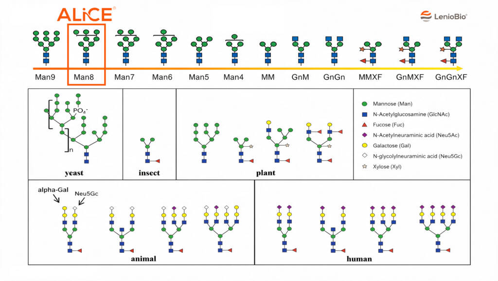
Two plant-specific glycan modifications can appear on ALiCE®-expressed proteins: α1,3-fucosylation on the core GlcNAc residue and β1,2-xylosylation. These are often present a very low levels and can be difficult to detect within regular assays. Both are characteristic of Nicotiana spp. expression systems generally — transient tobacco expression, stable BY-2 lines, and the BY-2 cell-free system alike — and distinguish plant-derived recombinant proteins from mammalian cell-derived proteins [3].
For research applications and most structural biology work, plant-specific glycosylation is not a practical concern: the glycan architecture does not alter the protein fold, and Man8 high-mannose glycans do not elicit significant responses in most in vitro assays. For vaccine antigen production, high-mannose glycans may be advantageous, since they are recognised by DC-SIGN and mannose receptors on dendritic cells and macrophages, potentially improving antigen uptake and presentation [4].
For therapeutic applications where the glycan directly determines function — IgG Fc effector function, FcRn-mediated half-life, complement activation — the plant-specific modifications warrant consideration. α1,3-fucose has been associated with IgE-mediated responses in some studies, though the clinical significance for biopharmaceuticals remains under investigation. Separately, the absence of the core α1,6-fucose found on CHO-derived antibodies, which reduces ADCC by impairing FcγRIII binding, means ALiCE®-produced antibodies may show enhanced ADCC potential — a potentially useful attribute where ADCC forms part of the mechanism of action.
There is already an FDA-approved therapeutic, PRX-102 (pegunigalsidase alfa), which is produced in the BY-2 cell line. This therapeutic enzyme has been developed for the treatment of Fabry disease,and is sold under the brand name Elfabrio. This supports the compatibility of BY-2 cell modifications with human patients.
The Route to Humanised Glycosylation
Two complementary approaches are in play. The first is host engineering: glycoengineered BY-2 cell lines with knockouts of the plant-specific α1,3-fucosyltransferase and β1,2-xylosyltransferase genes are under development [3]. Lysate produced from such lines would eliminate the plant-specific modifications, giving a cleaner high-mannose profile considerably closer to the Man8 glycoform mammalian cells produce under certain conditions.
The second exploits the open nature of the cell-free format. Because ALiCE® supports co-expression, glycan-modifying enzymes — GlcNAc transferases, galactosyltransferases, sialyltransferases — can in principle be expressed alongside the target protein to drive processing toward complex-type glycoforms within the reaction itself. Co-expression of OST variants and glycan-modifying enzymes has been identified as a route to homogeneous glycan profiles at scale [5]. This is an area of active investigation rather than a demonstrated capability.
The relative importance of N-glycosylation varies between protein classes. In all instances, a role for glycan-mediated folding and stability is acknowledged but very few functional roles have been described outside of the Fc receptor engagement for antibodies. To validate this further, LenioBio will be exploring additional therapeutic models as part of our InnoManContiMod project, co-funded by the European Union (https://www.leniobio.com/eu-grant-info/). Beyond classical antibodies, we will be producing novel, multispecific antibody formats and testing their efficacy against mammalian comparators. Vaccine antigens and growth factors are also included in the project scope and we look forward to sharing the results of these additional comparability studies in future updates.
Key Data
- Dominant N-glycan species: Man8 oligomannose, confirmed by LC-ESI-MS/MS
- Proteins characterised: glucose oxidase (5 sites), adalimumab (1 site), SARS-CoV-2 RBD (2 sites)
- High site occupancy across all characterised glycosites
- Reproducible glycoform distributions across multiple independent lysate batches
- Plant-specific modifications present: α1,3-fucose and β1,2-xylose, at low levels which may be difficult to detect in most proteins
- Partial complex-type processing observed at some sites (hybrid and GnGn species at RBD N343)
Frequently Asked Questions
What N-glycan structures does ALiCE® actually produce?
Predominantly oligomannose-type N-glycans with Man8 as the dominant species, confirmed by LC-ESI-MS/MS for glucose oxidase, adalimumab, and SARS-CoV-2 RBD. Plant-specific α1,3-fucosylation and β1,2-xylosylation are present at very low levels in most proteins, , and some sites show hybrid-type and GnGn species indicating partial complex-type processing. Site occupancy is high and profiles are reproducible across independent lysate batches.
How does ALiCE® glycosylation compare to CHO cell glycosylation?
CHO cells produce complex-type biantennary N-glycans with core α1,6-fucose — the G0F/G1F/G2F series on antibody CH2 domains. ALiCE® produces high-mannose Man8 profiles carrying low levels of plant-specific α1,3-fucose and β1,2-xylose. For research reagents and structural biology this distinction is usually immaterial. For therapeutic antibodies whose Fc effector function depends on complex-type glycans, the difference may be significant, depending on the final application and mode of action.
Can ALiCE®-produced glycoproteins be used for therapeutic development?
For applications where Man8-type glycosylation is acceptable or advantageous, yes. High-mannose glycans on vaccine antigens may improve uptake by mannose-binding lectins on antigen-presenting cells. For research reagents, structural biology targets, and functional assays, glycan type is rarely the limiting factor. For IgG therapeutics requiring complex-type glycans for FcγR engagement, further investigation is required to assess compatibility based on the final mode of action.
Why can’t most cell-free systems glycosylate proteins at all?
Most large-scale CFPS systems use E. coli extracts, which lack the eukaryotic secretory pathway entirely. Glycosylation requires an ER-resident OST complex to transfer the core oligosaccharide, a dolichol-linked precursor pool, glucosidases for trimming, and Golgi enzymes for further processing. None are present in prokaryotic lysates. ALiCE® is unusual among scalable CFPS platforms in retaining intact microsomes from the ER and Golgi that carry this machinery natively.
Is the glycosylation reproducible between batches of lysate?
Yes. Glycan profiles were analysed across multiple independent lysate batches and showed high reproducibility of glycoform distributions. The commercial release specification uses eYFP yield as a batch quality indicator, and the consistency observed in the characterisation studies is consistent with the controlled cell culture and lysate preparation process behind the commercial product.
Does high-mannose glycosylation affect protein stability or aggregation?
High-mannose glycans generally support correct folding and confer protease resistance in the same way complex-type glycans do, since the stabilising contribution comes largely from the core glycan and its interaction with the polypeptide surface. Where the glycan participates directly in a binding interface or in receptor-mediated clearance, the difference between high-mannose and complex-type becomes functionally relevant. Assessing this for a specific target requires direct comparison against a reference material.
References
[1] Zawada JF et al. Microscale to manufacturing scale-up of cell-free cytokine production. Biotechnol Bioeng. 2011;108(7):1570–1578. https://doi.org/10.1002/bit.23103
[2] Tinafar A, Jaenes K, Pardee K. Synthetic Biology Goes Cell-Free. BMC Biol. 2019;17:64. https://doi.org/10.1186/s12915-019-0685-x
[3] Das Gupta M et al. ALiCE®: A versatile, high yielding and scalable eukaryotic CFPS system. bioRxiv. 2022. https://doi.org/10.1101/2022.11.10.515920
[4] Hu YC, Kamat NP. Cell-free protein synthesis systems for vaccine design. Curr Opin Biotechnol. 2023;79:102888. https://doi.org/10.1016/j.copbio.2022.102888
[5] Buntru M et al. Plant-Derived Cell-Free Biofactories for the Production of Secondary Metabolites. Front Plant Sci. 2022;12:794999. https://doi.org/10.3389/fpls.2021.794999Funded by the European Union. Views and opinions expressed are however those of the author(s) only and do not necessarily reflect those of the European Union or the European Health and Digital Executive Agency. Neither the European Union nor the granting authority can be